RadioReport® Automatic AI is the further development of structured reporting.
We have mapped the thought processes of experienced radiologists into RadioReport® Automatic AI. The software guides you like a virtual interview to the final report – starting with / focused on anatomy instead of pathology. Instead of hundreds of templates, RadioReport® Automatic AI covers the entire range of MRI/CT indications with only 23 modules. The end result is a standardized report with machine-readable content ready for future AI applications.
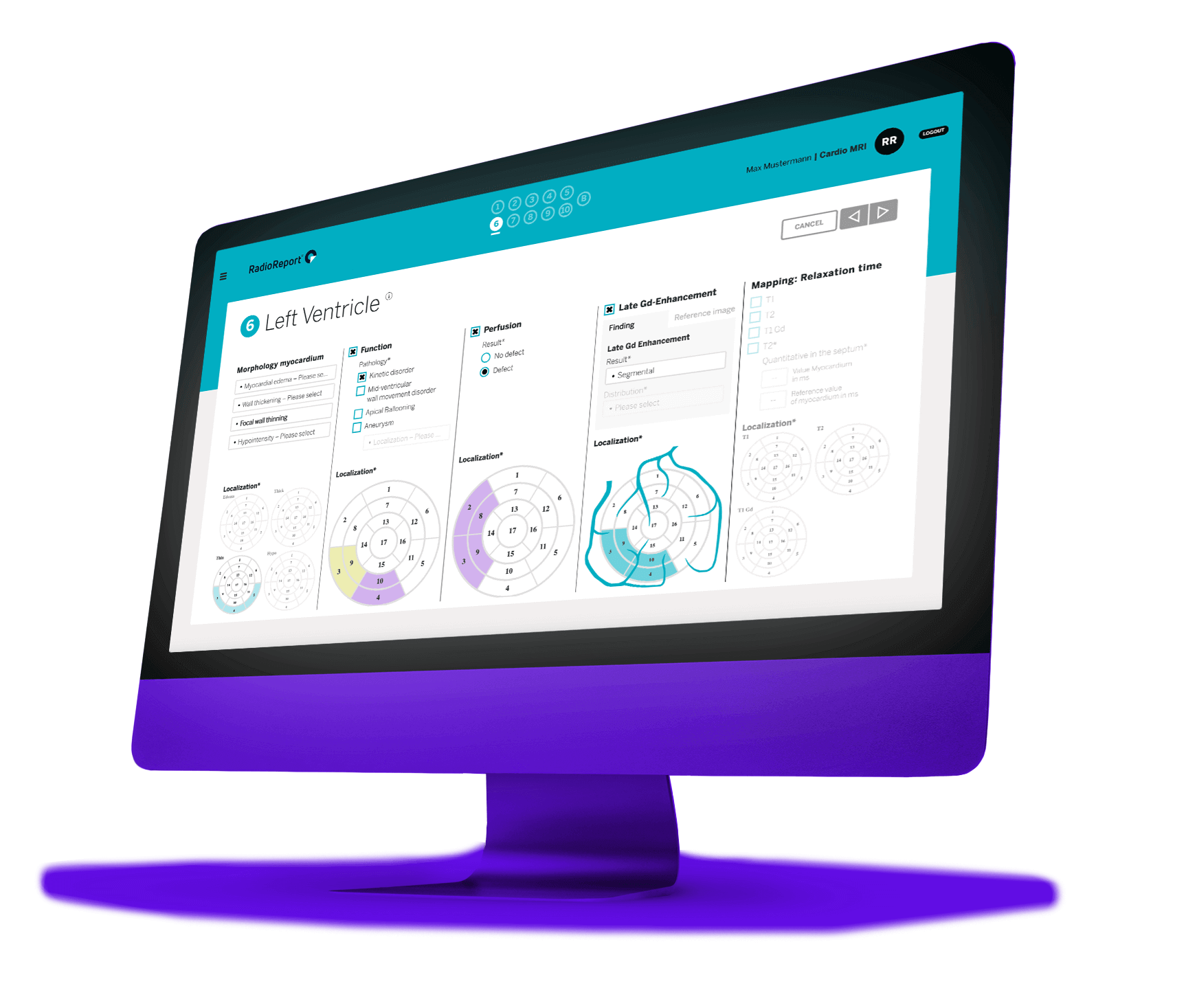
The human being is the focus of attention
Radiology is a high-tech specialty. Scanners are getting faster and faster while generating more and more data. What has hardly changed is reporting – the core activity of the radiologist. RadioReport® Automatic AI was developed to give people in the system room to maneuver and new opportunities for development.
From image to report in under 80 seconds!
With RadioReport® Automatic AI, you can achieve an enormous increase in efficiency.
With RadioReport® Automatic AI you achieve unprecedented efficiency. Complex report suggestions are generated with just a few mouse clicks. This gives you more time for image analysis, your patients or other operational activities.
With RadioReport® Automatic AI, you receive a high-quality, standardized, and easy-to-understand report suggestion containing relevant information for healthcare professionals. This ensures consistency and clarity for every radiologist or medical professional using RadioReport® Automatic AI in their decision-making process.
Since RadioReport® Automatic AI is multilingual, it can be used by an international network, allowing radiologists to offer their expertise and services worldwide.
From image to report in just a few steps
Exemplified by the “Knee MRI” module
RadioReport® Automatic AI enhances the consistency and clarity of reports. Mandatory fields help to avoid omissions and in-built plausibility checks prevent contradictions. This avoids inconsistencies between the findings and report.

Report up to
50% faster

Modules rather than templates

Anatomy rather
than pathology
Higher standards
of quality
Ready for
Big Data

Multilingual
Unlike other approaches to facilitate reporting in radiology, RadioReport® Automatic AI uses complete modules rather than templates. Since each module covers an entire range of indications, you can report the region studied holistically.
RadioReport® Automatic AI is driven by the anatomy, not the pathology. The software guides you like a virtual interview through the reporting process. The end result is a complete standardized report with machine-readable content.
These open up new business models for you. The value of the data generated by your reporting is considerable. It is measured as a benefit to medicine and is matched by a monetary equivalent. Data costs (but also earns) money.
Further topics
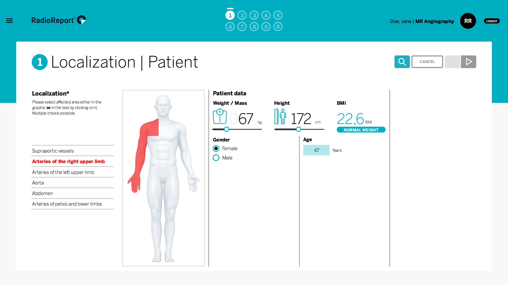
MR Angiograpy Module
The MR angiography module covers the full spectrum of MR angiography imaging findings. It helps you create accurate, standardized, and concise radiology reports for several vascular-related disorders.
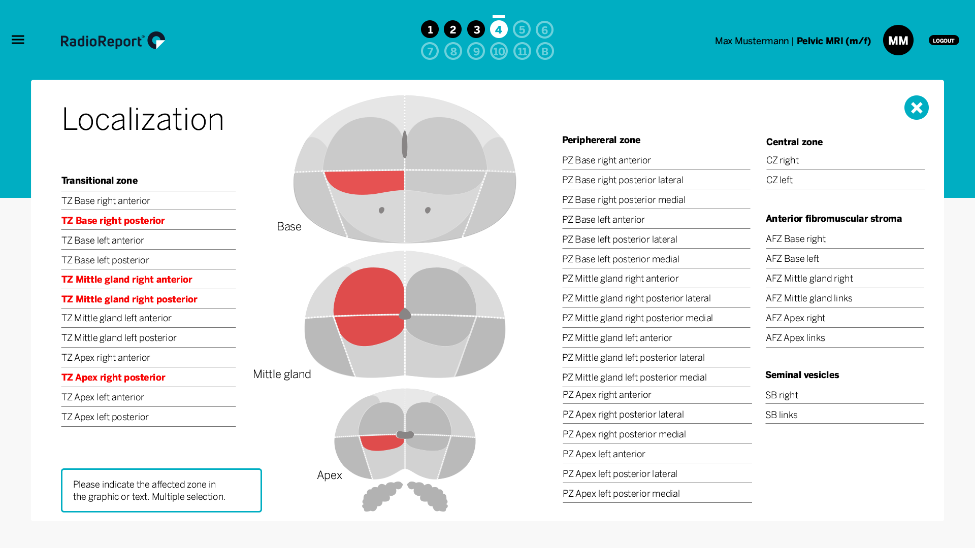
Pelvis MRI Module
This module covers the full spectrum of pelvis-MRI imaging findings. It provides high-quality, effective, and standardized radiology reports for several pelvis-related clinical indications.
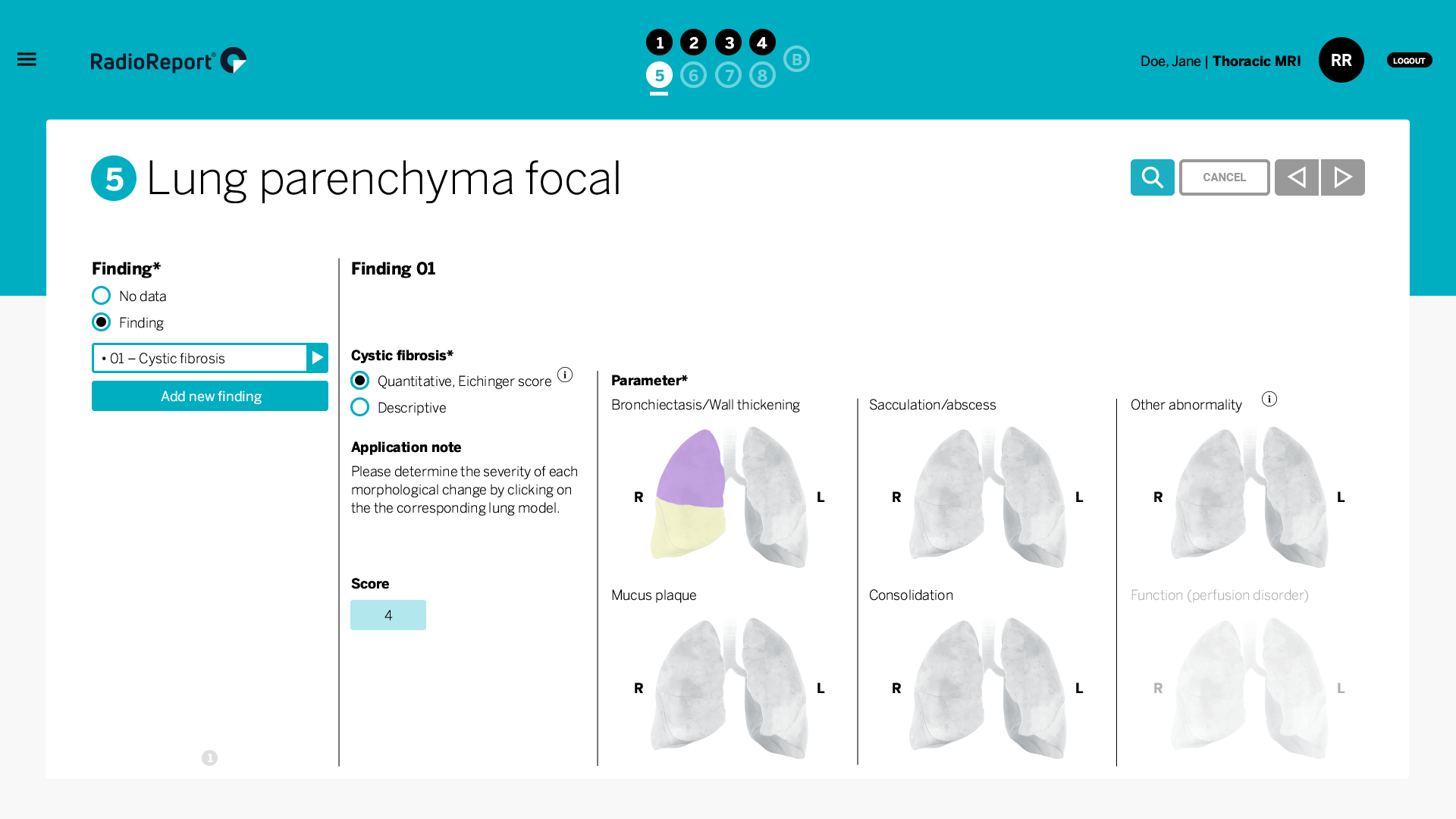
Chest MRI Module
Chest MRI was considered an infrequent radiological examination, but it is becoming a valuable routine diagnostic tool without exposure to radiation. This module helps you create accurate, standardized, and concise radiology reports for several chest-related indications.
