
Designed for
better reports.
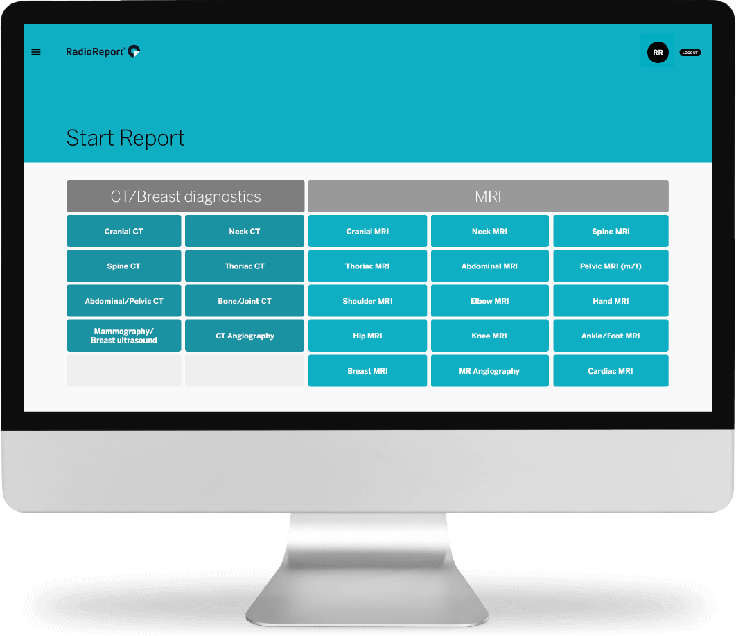
Anatomy, not pathology.
The User Experience (UX) and User Interface (UI) design from RadioReport® Automatic AI was developed in close collaboration with radiologists. What we learned: Reporting software has to begin with anatomy, not with pathology. A total of 23 modules for CT and MRI covers approximately 98% of all examinations in clinical practice.
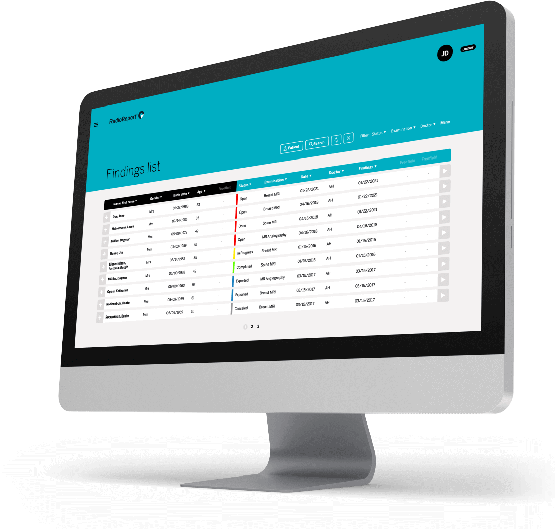
Where do you want to go today?
The control module of RadioReport® Automatic AI is the findings list – it offers a quick prioritized overview of current tasks. The design here focuses on clarity and function. Priorities are marked red and automatically sorted to the top.
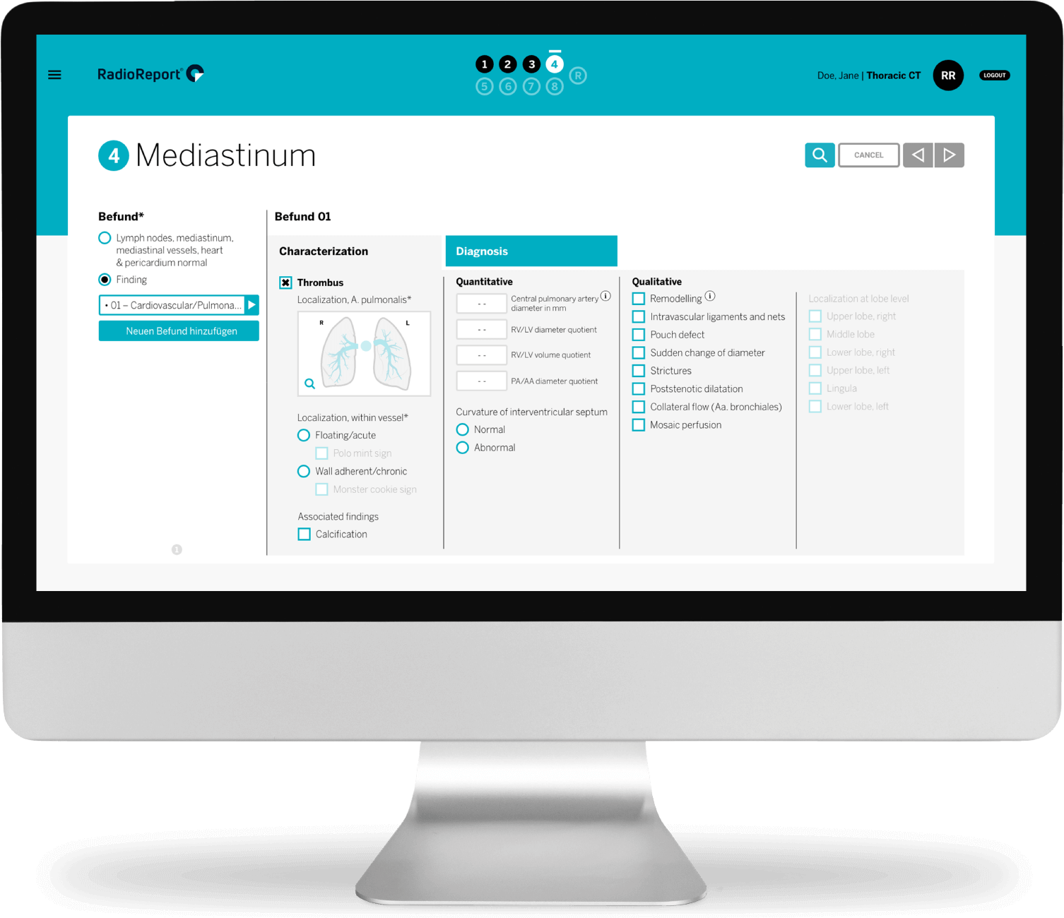
Form factor.
The core of RadioReport® Automatic AI are the forms that intuitively guide the user through the report. Each form deals with an anatomical or pathological aspect of a specific body region. Mandatory fields and inbuilt plausibility checks ensure that omissions are avoided.

Follow me.
The unique “guided reporting” approach of RadioReport® Automatic AI follows predefined steps, reflecting the workflow of experienced radiologists. Thus, “guided reporting” enables any user to report at a consistent quality and pace.

Skip dictation –
let RadioReport® Automatic AI do the talking.
Every box checked subsequently generates standardized text that appears in the final report.

Example of an automatically generated, standardized report suggestion.
“Finding of an acute pulmonary artery embolism. Evidence of a thrombus. Localization: segmental artery lateral middle lobe (S4), left pulmonary artery, segmental artery left apical lower lobe (S6), segmental artery left lateral lower lobe (S9).”
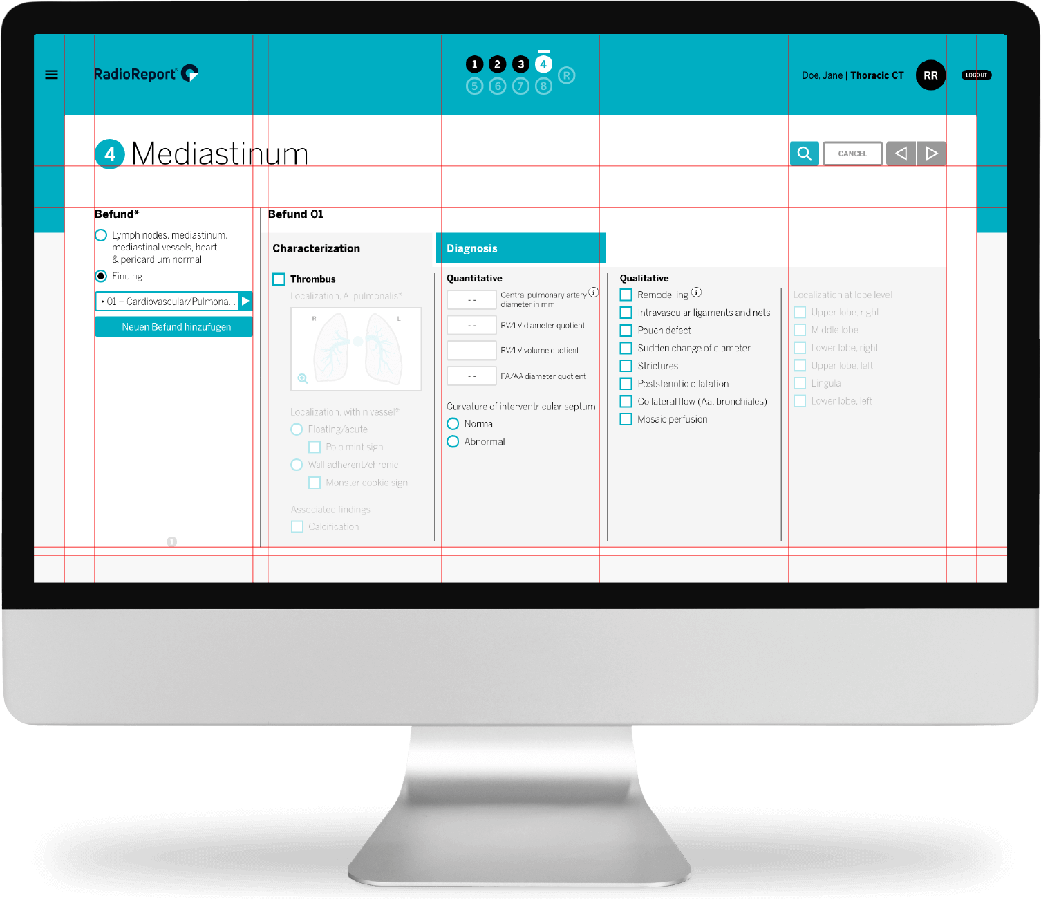
High five!
The five-column design was a key consideration during development of the software interface. The consistent implementation of this concept throughout RadioReport® Automatic AI provides quick orientation without forcing the user to scroll or search manually. As a result of this design, RadioReport® Automatic AI users significantly reduce their reporting time compared to conventional reporting methods.
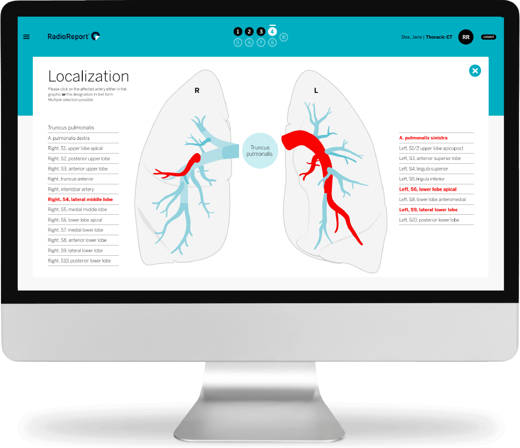
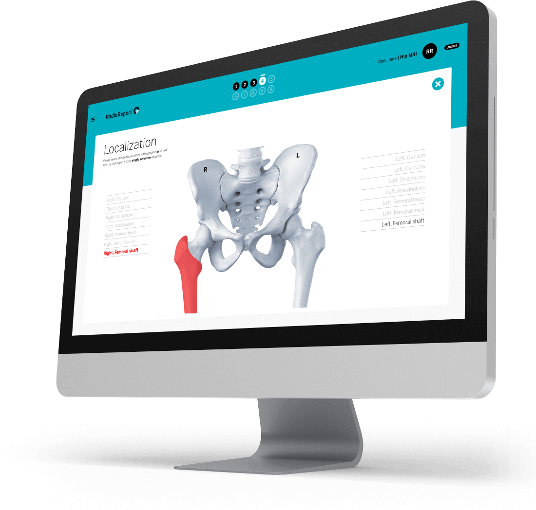
Localize with one click.
Visual localizers enable error-free data input and provide anatomical information as you go. No more need to refer to text books or web search. All localizers are concise graphic elements and serve as milestones throughout the reporting process. Working with them is intuitive and simple.
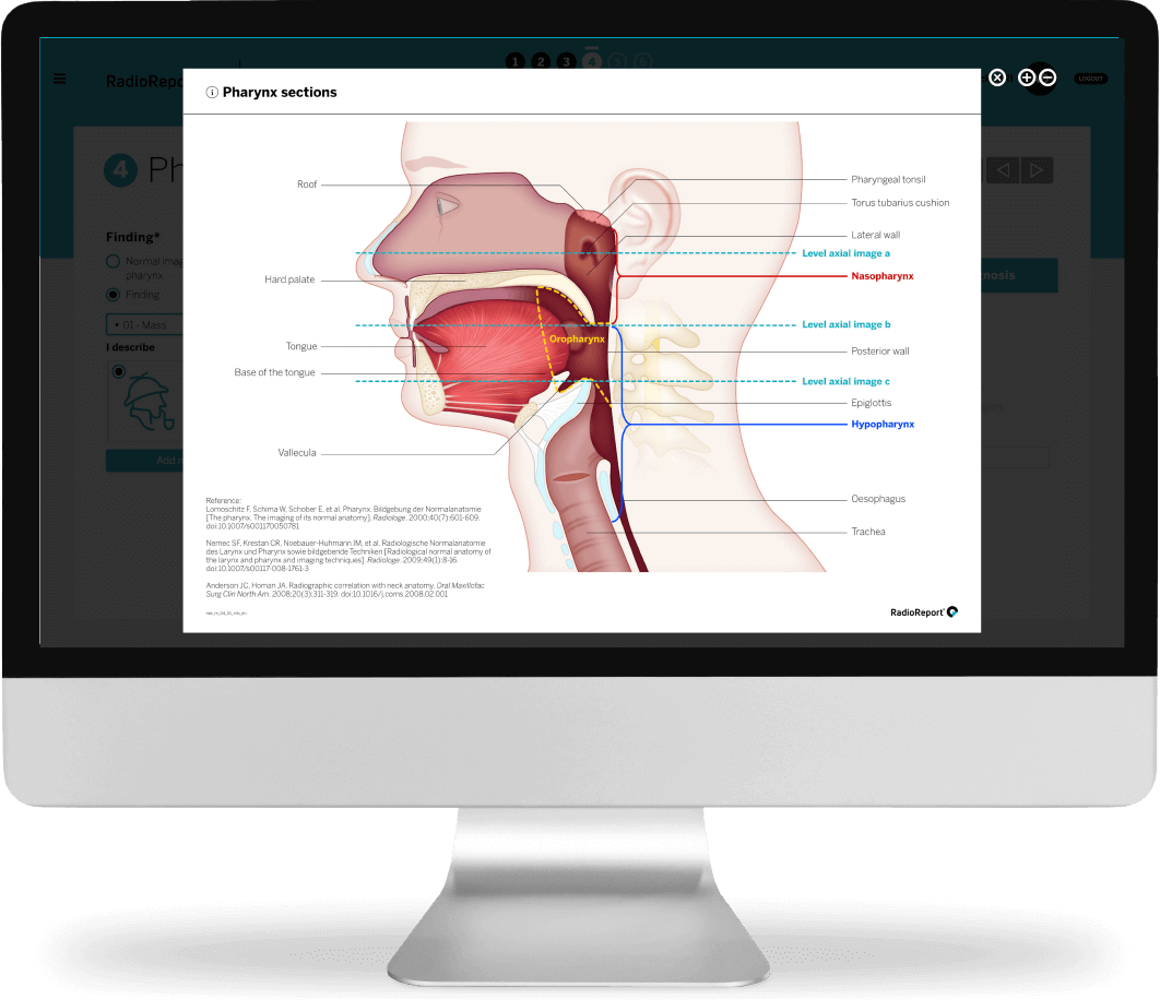
Expert knowledge always at hand.
Overlays provide information on anatomy, guidelines, et cetera and complement the diagnostic section, avoiding tedious searching in books, journals or the internet.
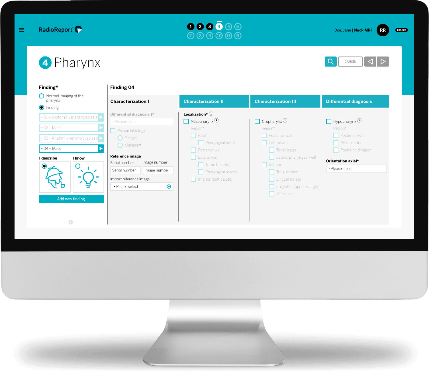
Unlimited findings.
With RadioReport® Automatic AI multiple pathologies are stored in the first column and remain visible throughout reporting. Sub-findings can be created, viewed, modified or deleted at any time.

Thanks, Sherlock!
The Sherlock and light bulb icons offer another feature: access to an individual analysis path or time-saving presets.
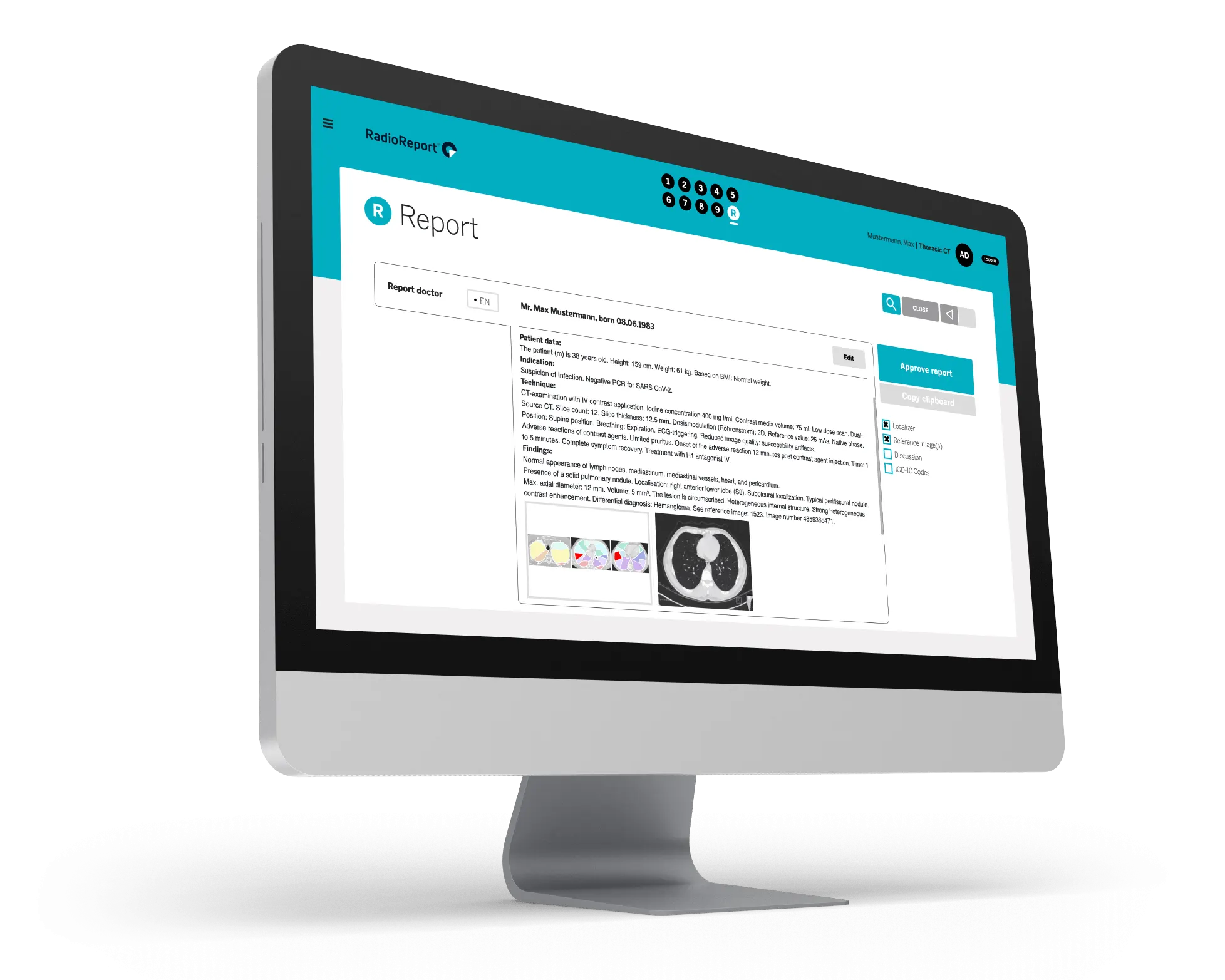
Experience the future
of structured reporting.
RadioReport® Automatic AI generates a complete report text: 100% digital, uniformly structured and fully machine-readable. Reference images and individual amendments can be included ad hoc. With one mouse click, reports can be generated in a number of languages.